9 December، 2025
Course entitled ” Introduction to medical imaging devices and medical image reading “

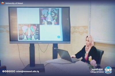
The Department of Medical Physics organized a course entitled (Introduction to medical imaging devices and medical image reading) the course was held on Sunday, November 30 at the seminar hall in the Department of medical physics under the supervision of Prof. Dr. Wahid Mahmoud Brahimi, dean of the College of Science, Prof. Dr. Hiyam Adel Ibrahim, and the follow-up of the head of the department Dr. Mahmoud Ahmed Mohammed Fakhri, the Department of Medical Physics organized a course entitled (Introduction to medical imaging devices and medical image reading). Shaima Talal Abdullah and Assistant L. Ayaz Rashid.
The course focused on understanding the medical imaging devices used in hospitals, and how to read the basic images of each device.
The course provides a clear explanation of the difference between the devices and why we use each of them.
During the course, the basic principles of medical imaging were explained such as contrast, accuracy, the principle of operation of each device, the physics of the device, and how to read the images resulting from a device. Then move on to display the following most important devices in a simplified form:
X-ray: identify how the image is formed and its uses in bone and chest examination.
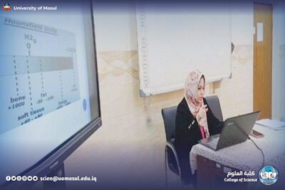
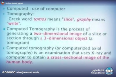
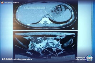
CT: an explanation of how to take multiple clips, the idea of the HU, and its role in imaging the brain, abdomen, and lung.
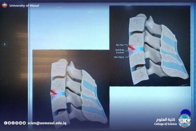
MRI: simplifying the principle of T1 and T2, understanding the high contrast of tissues, and its most important uses in the brain and spine.
PET: explain the idea of absorbing radioactive material and read the SUV for detecting tumors.
Mammography: simplifies the mechanism of detection of breast calcifications and lumps using low-dose radiography.
DXA: explanation of bone densitometry using dual-energy rays.
Government information and Communication Division
Tuesday, December 9, 2025